
Evolution of SPM for Biological Imaging Application Note

This article introduces the evolution of scanning probe microscopes in biological imaging
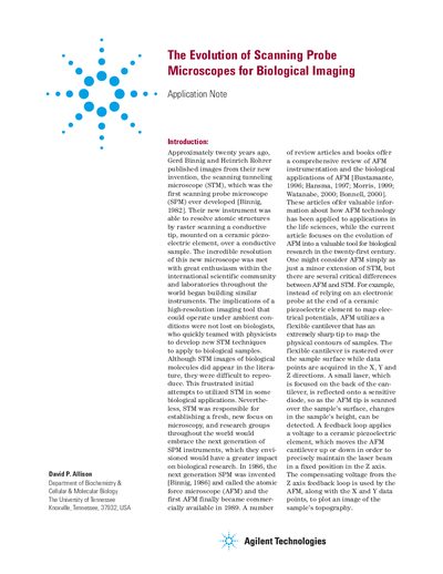

Evolution of SPM for Biological Imaging Application Note - cover page
Brand
Agilent
Document type
Application Note
Language
English
Pages
12
File format
PDF
File size
487 KB
Published
02 July, 2012
Updated
30 September, 2023
MD5 checksum
8B0594CED3036433D140DAADB4CF7BD1

About this manual

The document provides an overview of the history of scanning probe microscopy, starting with the Scanning Tunneling Microscope (STM) and evolving into the Atomic Force Microscope (AFM). It highlights how AFM overcomes limitations of STM by being able to image non-conductive materials like biological samples.

What's inside

  • Introduction
  • Evolution of the AFM
  • Imaging with AFM: Contact Mode
  • Imaging with AFM: Dynamic or AC AFM

Frequently asked questions

What types of biological samples can AFM image that STM cannot?

AFM is able to image non-conductive or insulating materials, such as proteins, DNA, and living cells.

Why must biological samples be firmly affixed when using contact mode AFM?

Physical interactions in contact mode create significant lateral force, so samples must be firmly affixed to an atomically flat substrate.

How does AC AFM avoid the sample degradation seen in contact mode?

AC AFM encounters negligible lateral forces, making it a gentler technique that prevents severe sample degradation.

Download PDF (487 KB)

More Agilent Manuals


View all Agilent manuals →