
Peak 2 was correctly identified to contain only tyrosine, peak 3 contained neither tyrosine nor tryptophan and peaks 4 and 6 contained only tryptophan (figure 1). Second derivative spectra can be used to emphasize small spectral differences in similar molecules. This is especially useful for identifying tryptophan and tyrosine residues in peptides (figure 2).
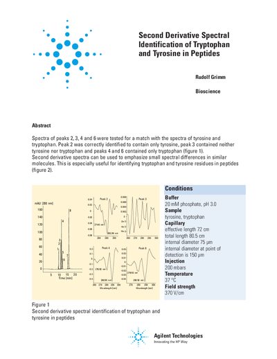
This document presents a study on identifying tryptophan and tyrosine residues within peptide samples. The method utilizes second derivative spectral analysis to distinguish between similar molecules, specifically highlighting peaks corresponding to these amino acids.
The application was performed using an Agilent Capillary Electrophoresis system with specific conditions including a 20 mM phosphate buffer at pH 3.0 and a temperature of 37 °C. The results demonstrate successful identification where peak 2 contained only tyrosine, while peaks 4 and 6 contained only tryptophan.
| Buffer composition | 20 mM phosphate, pH 3.0 |
|---|---|
| Sample type | tyrosine, trytosine |
| Capillary effective length | 72 cm |
| Capillary total length | 80.5 cm |
| Injection pressure | 200 mbars |
| Temperature | 37 °C |
| Field strength | 370 V/cm |
They are used to emphasize small spectral differences in similar molecules, which helps identify tryptophan and tyrosine residues.
Peak 2 was correctly identified to contain only tyrosine.