
This application note introduces the basic principle and applications of fluorescence microscopy, as well as the features and advantages of Agilent fluorescence microscopes.
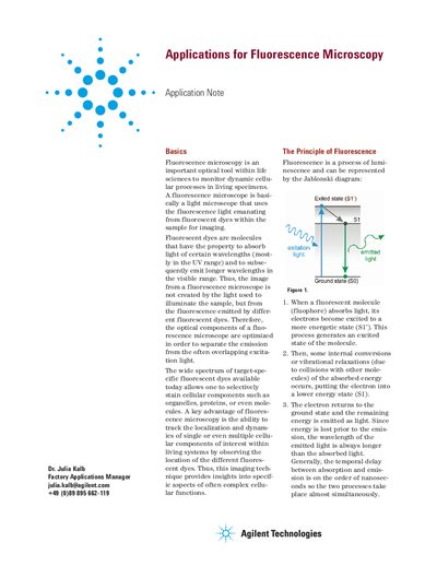
Fluorescence microscopy is an optical tool used in life sciences to monitor dynamic cellular processes in living specimens. It utilizes fluorescent dyes that absorb light at specific wavelengths and emit longer wavelengths in the visible range, allowing for the imaging of specific cellular components like organelles and proteins. The document explains the Jablonski diagram to illustrate the process of excitation and emission, noting that the emitted light has a longer wavelength than the absorbed light due to energy loss during internal conversions.
It uses fluorescence light from dyes within a sample to image dynamic cellular processes in living specimens.
The image is created from fluorescence emitted by different dyes rather than the light used to illuminate the sample.
It allows tracking the localization and dynamics of single or multiple cellular components within living systems.