
This document presents an interactive Matlab® interface for manual dermoscopic image analysis. This interface allows performing the manual segmentation of dermoscopic images and storing the result of segmentation. Its main functionalities are image upload and display, manual segmentation, boundary reshaping and storage of segmented image.
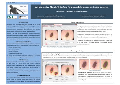
This document describes an interactive MATLAB interface for manual segmentation of dermoscopic images. It is intended to help build a ground-truth database of manually segmented images for validating computer-aided diagnosis algorithms.
The interface supports image upload and display, manual segmentation, boundary reshaping, and storage of segmented images. It was implemented in MATLAB 7.9.0 R2009b and includes pointwise and local boundary reshaping methods.
| MATLAB environment | 7.9.0 R2009b |
|---|
It is used for manual segmentation of dermoscopic images and storing the segmentation results.
The user clicks and drags control points to their new positions.
A binary mask of the initial contour is created, then morphological filtering is applied.