
This document describes the usage and principle of the JC-1 Apoptosis Detection Kit for measuring mitochondrial membrane potential. The kit utilizes JC-1 as a fluorescent dye to indicate the mitochondrial membrane potential. It can be used to detect the changes in mitochondrial membrane potential before and after cell apoptosis. The changes in mitochondrial membrane potential can be observed using fluorescence microscopy or flow cytometry, allowing for the determination of cell apoptosis.
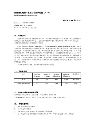
The Kaiji JC-1 Apoptosis Detection Kit is designed to detect changes in mitochondrial transmembrane potential (Δψ) in cells, tissues, or purified mitochondria. It utilizes the JC-1 cationic lipid fluorescent dye, which exists as monomers and polymers depending on membrane potential. In healthy cells with high potential, JC-1 forms red-fluorescing polymers within mitochondria; in apoptotic cells where potential collapses, it releases as green-fluorescing monomers into the cytoplasm. This manual is written in Chinese.
| Storage Temperature (Kit) | -20℃ |
|---|---|
| Excitation Wavelength (Microscopy Monomer) | 490nm |
| Emission Wavelength (Microscopy Monomer) | 530nm |
| Excitation Wavelength (Flow Cytometry) | 488 nm |
| Emission Wavelength (Flow Cytometry Monomer) | 530 nm |
The manual recommends collecting a cell amount of 1×10^6 per test.
JC-1 must be stored at -20℃ in the dark and used while avoiding light exposure.
A decrease in mitochondrial membrane potential results in green fluorescence (monomer) and reduced red fluorescence intensity.