
Manual for Making Cytological Specimens of Body Cavity Fluid

This document introduces the manual for making cytological specimens of body cavity fluid.
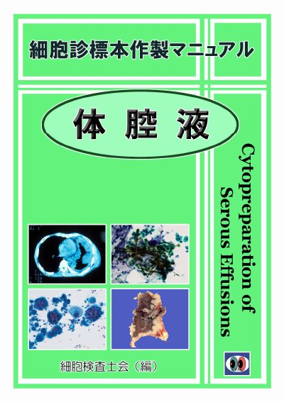

Manual for Making Cytological Specimens of Body Cavity Fluid - cover page
Document type
Other
Language
Japanese
Pages
24
File format
PDF
File size
2.4 MB
Published
18 November, 2023
Updated
18 November, 2023
MD5 checksum
EA2DFABFD3D74DCF645B6E8B0BDBA553

About this manual

This manual gives detailed guidance for preparing cytological specimens from body cavity fluid, from specimen collection and macroscopic assessment through processing of bloody specimens, centrifugation, and smear preparation.

It also covers methods to reduce cell detachment, recommended slide counts, fixation, body cavity washing fluid specimens, Giemsa and May-Giemsa staining, immunocytochemical staining, cell block preparation, and cell transfer. This manual is written in Japanese.

What's inside

  • Handling flowchart and specimen collection
  • Processing of bloody specimens
  • Centrifugation speed and residual cells
  • Smear methods and cell detachment prevention
  • Fixation and slide counts
  • Body cavity washing fluid specimens
  • Giemsa and May-Giemsa staining
  • Immunocytochemical staining
  • Cell block preparation
  • Cell transfer method

Frequently asked questions

How many slides are recommended at minimum?

At least 3 wet-fixed slides and 1 dry slide are recommended.

What centrifuge speed is used to collect cells?

Centrifugation at about 3000 rpm for about 3 minutes is used to collect cells.

How long can specimens be refrigerated?

Even after 48 hours of refrigeration, cytological diagnosis is often still possible.

What hemolysis agents are used for bloody specimens?

Hemolysis agents include 0.9% ammonium chloride solution and 1.2% ammonium oxalate solution.

Download PDF (2.4 MB)

Also Manuals