
This document introduces the use of Neodent Guided Surgery system, including preoperative preparation, intraoperative operation and postoperative care
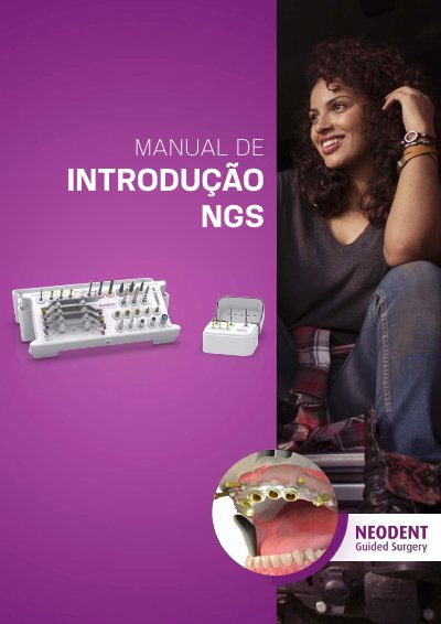
The Neodent Guided Surgery (NGS) system assists surgeons in performing implant treatments based on pre-planning using advanced software. The system aims to improve predictability, aesthetics, and patient comfort while reducing surgical time and trauma. It supports various rehabilitation scenarios, including partial and total rehabilitation, and offers specific kits for different implant types and clinical situations.
The manual details the necessary steps for the surgical procedure, starting with diagnosis and Cone Beam Tomography. It explains the creation of the tomographic guide and the double scanning technique required for accurate data acquisition. Furthermore, it describes the preparation of STL files, the workflow with coDiagnostiXTM software, and the fabrication of the surgical guide. Specific sections cover the use of various surgical kits (Titamax, Alvim, Facility, Start) and the installation of implants using compatible instruments. This manual is written in Portuguese.
| Minimum FOV diameter | 8 cm |
|---|---|
| Tomographic guide material | Acrylic |
| Guide marking depth | 1 mm |
| Dental separation minimum | 2.0 mm |
Yes, the professional should undergo training through a Guided Surgery NGS course offered by partner educational institutions.
The separation of the teeth must be at least 2.0 mm.
The Field of View must be a minimum of 8 cm in diameter.
The guide must have a minimum thickness of 2 mm.