
This article introduces the principles and applications of biopotential electrode sensors in electrocardiography (ECG), electromyography (EMG), and electroencephalography (EEG) systems. Biopotential electrodes can monitor heart, muscle, and brain activity by measuring electric potentials on the surface of living tissue. These electrodes sense ion distribution on the tissue surface and convert ion current to electron current. An electrolyte solution/gel is applied to one side of the electrode in contact with the tissue, and a chemical reaction occurs at the interface between the electrolyte and the electrode. Ions are discharged into the electrolyte, while electrons carry charge through the lead wires. This technology finds wide applications in medical monitoring and diagnostics.
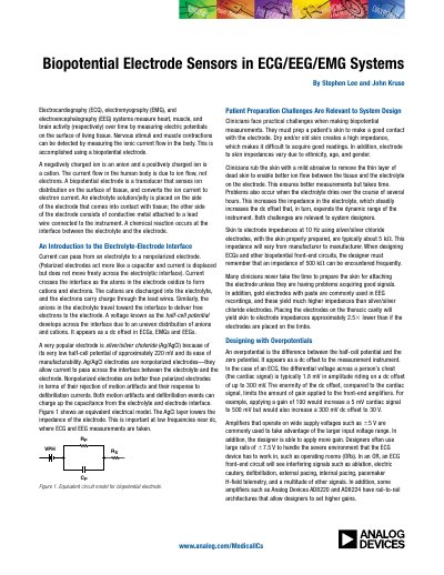
The document provides an introduction to biopotential electrode sensors used in electrocardiography (ECG), electromyography (EMG), and electroencephalography (EEG) systems. It details the mechanism of ion-to-electron current conversion at the electrolyte-electrode interface, highlighting the characteristics of silver/silver chloride electrodes.
The impedance is typically about 5 kΩ.
A voltage known as the half-cell potential develops across the interface due to an uneven distribution of anions and cations.
JFET input op amps such as the AD8625/AD8626/AD8627 and AD8641/AD8642/AD8643 have input bias currents of less than 1 pA.