
This document introduces the use of Agilent nScope eMRI for the imaging of mouse heart. It is suitable for novice and expert cardiac researchers.
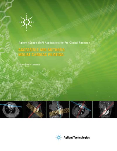
This document outlines the procedure for performing mouse cardiac piloting using the Agilent nScope eMRI system. It is designed to assist both novice and expert cardiac researchers in acquiring sophisticated images of mouse cardiac axes through simple, precise positioning of double oblique planes. The guide details the use of the Agilent Cardiac Pack, which features pre-selected, laboratory-tested protocols to simplify image acquisition for first-time and sophisticated users.
The instructions cover the specific steps required to obtain short-axis and long-axis views of the mouse heart. Users are guided through acquiring scout scans, prescribing oblique coronal and sagittal slices, and orienting stacks of slices perpendicular to the ventricular axis. The document also notes that these instructions are valid for all field strengths and RF coil configurations, provided a triggering signal synchronous with the ECG is supplied.
| Acquisition time per slice | ~ 4's (2 averages) |
|---|---|
| Heart rate range | 120-130 ms |
| Matrix size | 128 x 128 (zero filled to 256 x 256) |
| Slice thickness | 1 mm |
Yes, these instructions for mouse cardiac piloting methodology are valid for all field strengths and all RF coil configurations.
A triggering signal synchronous with the ECG of the animal must be provided to the scanner by an appropriate monitoring system.